🔬 Biology · Class 11 · NEET
Locomotion and Movement - Practice Questions with Answers
75 free MCQs on Locomotion and Movement, each with its own worked answer and explanation. Types of movement, muscle contraction, skeletal system, joints, and disorders. Important for Class 11 and NEET.
Take the timed Locomotion and Movement chapterwise test →75 practice questions on Locomotion and Movement, sorted Easy → Hard. Try each one first, then open its answer page for the worked explanation. Want the full theory first? Read the Locomotion and Movement notes.
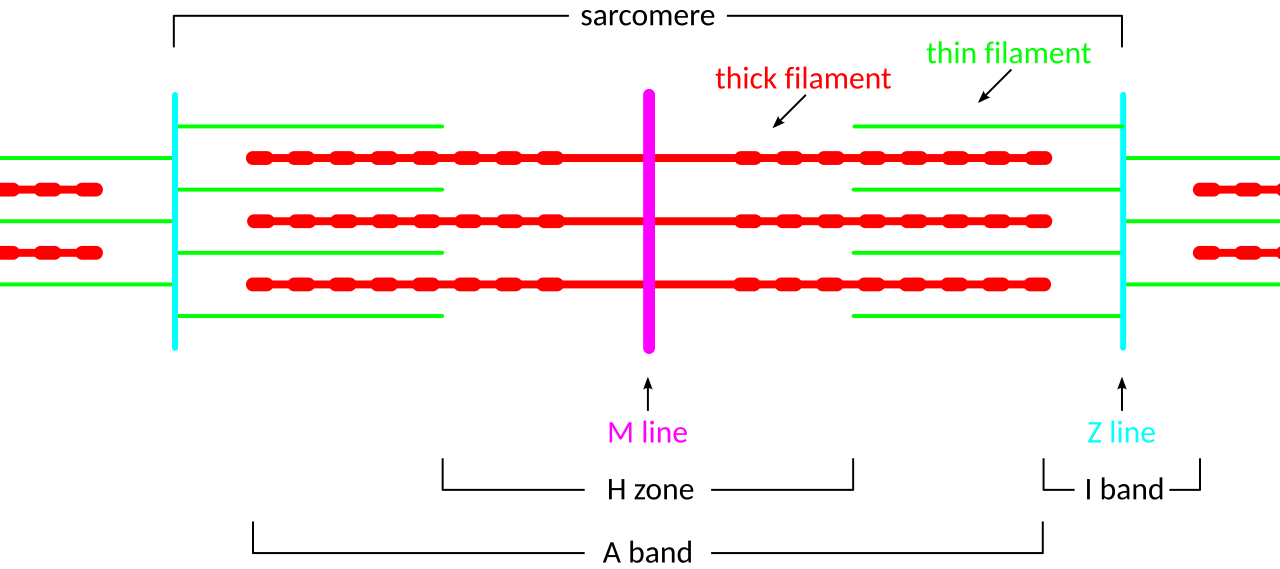
A sarcomere (Z line to Z line): during contraction the I band and H zone shorten as thin filaments slide over thick, while the A band stays the same length. Image: SlothMcCarty, CC BY-SA 3.0, via Wikimedia Commons.
Easy - 25 questions
Q4.
The immovable joints that join the bones of the skull are called:
- A Hinge joints
- B Pivot joints
- C Gliding joints
- D Sutures
Show answer & explanation →
Q5.
The muscle found in the walls of internal organs, working without conscious control, is:
- A Smooth muscle
- B Skeletal muscle
- C Cardiac muscle
- D Striated muscle
Show answer & explanation →
Q11.
What is the role of calcium ions in muscle contraction?
- A Provide chemical energy by being hydrolyzed directly at the myosin head region overall
- B Bind to troponin, causing tropomyosin to move and expose actin binding sites for myosin
- C Transport oxygen from nearby capillaries directly into the muscle fibre tissue in most cases
- D Form the actual cross-bridge structure by binding directly to actin filaments under typical conditions
Show answer & explanation →
Q12.
The axial skeleton consists of
- A Limb bones of the arms and legs mainly
- B Skull, vertebral column, and ribcage (80 bones)
- C Mainly the skull and its associated facial bones
- D Pectoral and pelvic girdles that attach the limbs
Show answer & explanation →
Q13.
What is osteoporosis?
- A A bacterial infection that spreads through the bone marrow cavity over time
- B Decreased bone density making bones fragile and prone to fracture
- C Excessive new bone growth causing abnormal thickening of the skeleton
- D Inflammation of the synovial joints and surrounding cartilage tissue
Show answer & explanation →
Q14.
What is the ball and socket joint? Give an example.
- A Allows movement mainly in one plane, as seen at the knee
- B Allows movement in all planes - hip and shoulder joints
- C Allows rotation mainly around a single axis, as at the atlas-axis
- D Allows almost no movement, as at the sutures in the skull
Show answer & explanation →
Q15.
What is the pivot joint? Give an example.
- A Allows simple bending and straightening, as commonly seen at the elbow joint according to standard textbooks
- B Allows rotation only - atlas-axis joint (head rotation), radioulnar joint (forearm rotation)
- C Allows movement in all possible directions, as at the ball-and-socket shoulder joint in general practice
- D Allows mainly side-to-side gliding movement, as between adjacent carpal bones as frequently described
Show answer & explanation →
Q16.
Which type of muscle is found in the heart?
- A Skeletal muscle
- B Smooth muscle
- C Cardiac muscle
- D Both skeletal and smooth
Show answer & explanation →
Q17.
What is rigor mortis?
- A A general relaxation of skeletal muscles caused by sudden calcium efflux shortly after death in most textbook accounts
- B Stiffening of muscles after death due to lack of ATP preventing myosin-actin cross-bridge detachment
- C A genetic muscle disease typically caused by a mutation in the dystrophin gene itself during normal conditions
- D Normal muscle fatigue resulting from lactic acid buildup during prolonged heavy exercise as generally observed
Show answer & explanation →
Q18.
What does the vertebral column protect?
- A Heart, cushioning it from external impact
- B Spinal cord and nerve roots
- C Lungs, by forming a rigid bony cage around them
- D Brain, by enclosing it within the cranial cavity
Show answer & explanation →
Q19.
What is the function of synovial fluid?
- A Carry nutrients directly into compact bone tissue
- B Lubricate the joint, reduce friction, and absorb shock
- C Form new cartilage at the articular surface
- D Attach muscle fibres firmly to the periosteum of bone
Show answer & explanation →
Q22.
Voluntary muscles are also called
- A Smooth muscles
- B Cardiac muscles
- C Skeletal muscles
- D Involuntary muscles
Show answer & explanation →
Q23.
What is arthritis?
- A A genetic muscle disease that causes slow, progressive muscle weakness over time
- B Inflammation or degeneration of joints causing pain and stiffness
- C A fracture of one of the long bones, usually caused by trauma or a fall
- D A nerve disease that specifically affects motor neuron signaling pathways
Show answer & explanation →
Q24.
The human vertebral column has how many vertebrae?
- A 26 (7C + 12T + 5L + 5S + 4Coccyx fused)
- B 22
- C 30
- D 33 (7C + 12T + 5L + 5S + 4Coccyx unfused)
Show answer & explanation →
Q25.
What is the main mineral that gives bones their hardness?
- A Iron, stored mainly in the bone marrow
- B Sodium, concentrated in the extracellular matrix
- C Calcium phosphate (hydroxyapatite)
- D Magnesium, bound loosely within collagen fibres
Show answer & explanation →
Medium - 25 questions
Q26.
During muscle contraction, the band whose length stays unchanged is the:
- A I band
- B A band
- C H zone
- D Whole sarcomere
Show answer & explanation →
Q27.
The central lighter region of the A band, having only thick filaments, is the:
- A I band
- B Z line
- C H zone
- D M line
Show answer & explanation →
Q28.
Calcium ions needed for muscle contraction are stored in the:
- A Golgi apparatus
- B Cell nucleus
- C Mitochondrial matrix
- D Sarcoplasmic reticulum
Show answer & explanation →
Q30.
The neurotransmitter released at the neuromuscular junction is:
- A Adrenaline
- B Acetylcholine
- C Dopamine
- D Serotonin
Show answer & explanation →
Q31.
Explain the sliding filament theory of muscle contraction.
- A Muscle contracts by folding the thin actin filaments into tightly compacted coils along the sarcomere structure as generally observed in typical laboratory settings
- B Thin actin filaments slide over thick myosin filaments using cross-bridge cycling powered by ATP; the sarcomere shortens while filament lengths stay constant
- C Myosin filaments physically shorten in their overall structural length during the course of muscle contraction itself under usual circumstances according to most researchers
- D Both the thin actin and thick myosin filaments shorten together by a roughly equal amount during contraction in the majority of cases studied as widely reported
Show answer & explanation →
Q32.
What changes occur in the sarcomere during muscle contraction?
- A The A band shortens noticeably while the I band stays mostly unchanged throughout contraction
- B I band and H zone decrease; A band stays same; Z discs come closer together
- C All visible bands of the sarcomere shorten by a roughly equal amount during contraction
- D Mainly the H zone changes noticeably, with little movement of the Z discs
Show answer & explanation →
Q33.
What is the neuromuscular junction and how does it transmit signals?
- A A direct, uninterrupted electrical connection that permanently couples the nerve cell membrane to the muscle cell membrane surface across the gap in standard practice under most conditions encountered
- B Synapse between motor neuron and muscle; acetylcholine released from nerve terminal binds nicotinic receptors on motor end plate, generating end-plate potential that triggers action potential in muscle
- C A thick myelin sheath layer that passively covers much of the outer surface area of the muscle fibre membrane in most vertebrate species as frequently observed in practice in many documented cases
- D A specialized gap junction structure that links together two separate adjacent skeletal muscle cell membranes electrically and chemically according to conventional understanding in routine practice
Show answer & explanation →
Q34.
What is muscle fatigue and what causes it?
- A Fatigue that is sometimes thought to be caused mainly by a temporary shortage of available oxygen reaching the muscle fibre overall in most cases under typical conditions
- B Inability to maintain force due to: depletion of ATP and creatine phosphate, lactic acid accumulation, ion imbalances (K+ accumulates outside cell), and glycogen depletion
- C Fatigue that is sometimes thought to be caused mainly by the gradual, ongoing accumulation of lactic acid within the fibre according to standard textbooks in general practice
- D Fatigue that is sometimes considered largely psychological in nature, with little underlying physiological basis involved as frequently described in most textbook accounts
Show answer & explanation →
Q35.
What is the difference between fast-twitch and slow-twitch muscle fibres?
- A Both fibre types are sometimes thought to be structurally, biochemically, and metabolically quite similar to one another in most measurable ways during normal conditions as generally observed
- B Fast-twitch (Type II): large, fast, powerful, fatigue quickly, anaerobic; Slow-twitch (Type I): small, slow, fatigue resistant, aerobic, more mitochondria, more myoglobin (red)
- C Fast-twitch fibres are sometimes mistakenly thought to occur mainly within cardiac muscle tissue, rarely elsewhere in the body in typical laboratory settings under usual circumstances
- D Slow-twitch fibres are sometimes mistakenly thought to be specialized for generating short, explosive bursts of sprinting power according to most researchers in the majority of cases studied
Show answer & explanation →
Q36.
What is the role of troponin and tropomyosin in muscle regulation?
- A Both of these proteins are sometimes thought to be mainly structural support elements, with little regulatory signaling role in the cell as widely reported in standard practice
- B Tropomyosin blocks myosin binding sites on actin at rest; troponin (TnC, TnI, TnT complex) senses Ca<sup>2+</sup>; Ca<sup>2+</sup> binding to TnC moves tropomyosin away, exposing binding sites
- C Troponin is sometimes mistakenly thought to single-handedly supply the chemical energy required for cross-bridge cycling under most conditions encountered as frequently observed in practice
- D Tropomyosin is sometimes mistakenly described as forming the actual structural cross-bridge linking actin and myosin filaments in many documented cases according to conventional understanding
Show answer & explanation →
Q37.
What is the structure of a typical synovial joint?
- A A joint type that largely lacks any fluid-filled cavity between its two articulating bone surfaces, unlike most movable joints in routine practice overall in most cases under typical conditions
- B Articular cartilage covers bone ends; synovial cavity contains synovial fluid; synovial membrane lines the joint capsule; fibrous joint capsule and ligaments stabilize the joint
- C Two bones that are permanently fused directly together by largely ossified, rigid connective tissue with little movement possible according to standard textbooks in general practice
- D Mainly a thin layer of articular cartilage is present at this joint, with very little synovial fluid contained inside the cavity as frequently described in most textbook accounts
Show answer & explanation →
Q38.
What is muscular dystrophy?
- A A chronic, slowly progressive degenerative disease that mainly affects the synovial joints throughout the body during normal conditions as generally observed
- B A group of genetic diseases causing progressive muscle weakness and degeneration; most common is Duchenne MD caused by dystrophin gene mutation on X chromosome
- C A disease that is sometimes thought to be caused mainly by a long-term, chronic dietary deficiency of vitamin D in typical laboratory settings under usual circumstances
- D An autoimmune disorder in which the body's own circulating antibodies are sometimes thought to attack healthy muscle fibres according to most researchers
Show answer & explanation →
Q39.
What is the role of ATP in muscle relaxation (not contraction)?
- A ATP is sometimes thought to play little functional role during the relaxation phase that follows muscle contraction in the majority of cases studied as widely reported
- B ATP is required for: Ca<sup>2+</sup> pump (SERCA) to return Ca<sup>2+</sup> to SR; myosin head detachment from actin after power stroke; active transport of ions to restore resting potential
- C ATP molecules are sometimes thought to be consumed mainly during the active contraction phase, rarely during relaxation in standard practice under most conditions encountered
- D Mainly ADP molecules, rather than ATP molecules, are sometimes thought to bring about muscle fibre relaxation as frequently observed in practice in many documented cases
Show answer & explanation →
Q40.
What is tendinitis and how is it caused?
- A A localized bacterial infection that gradually spreads throughout the protective fibrous sheath surrounding a particular tendon
- B Inflammation of tendons usually from repetitive strain or overuse; common sites: rotator cuff, Achilles tendon, patellar tendon, lateral epicondyle
- C A slow, chronic degenerative disease process that specifically and primarily affects the long bones found within the limb
- D Inflammation that occurs specifically and only within the fibrous ligaments themselves, rather than within the tendons connecting muscle to bone
Show answer & explanation →
Q41.
What is the difference between isometric and isotonic muscle contractions?
- A Both of these terms are sometimes thought to describe the same single underlying type of muscle contraction in any muscle according to conventional understanding in routine practice
- B Isometric: muscle generates force but does not change length (holding weight); isotonic: muscle changes length while maintaining relatively constant tension (lifting weight)
- C Isometric contraction is sometimes thought to usually involve visible joint movement, unlike isotonic contraction movement overall in most cases under typical conditions
- D Mainly skeletal muscle fibres are thought to be capable of undergoing a true isotonic type of contraction according to standard textbooks in general practice as frequently described
Show answer & explanation →
Q42.
What are the red and white fibres of skeletal muscle based on myoglobin content?
- A Red muscle fibres are sometimes mistakenly classified as fast-twitch while white fibres are instead mistakenly classified as slow-twitch in most textbook accounts
- B Red fibres (high myoglobin, slow-twitch, aerobic) appear red due to high myoglobin content; white fibres (low myoglobin, fast-twitch, anaerobic) appear pale
- C Both of these fibre types are sometimes thought to contain a roughly equal concentration of the oxygen-binding protein myoglobin in their cytoplasm during normal conditions
- D White muscle fibres are sometimes mistakenly thought to be the single most predominant fibre type found distributed throughout the entire heart wall as generally observed
Show answer & explanation →
Q43.
What is the pectoral girdle?
- A The pair of large hip bones that together form the structural base of the pelvis and lower limb attachment
- B Shoulder girdle consisting of clavicle and scapula on each side; connects upper limbs to axial skeleton
- C The sternum bone alone, existing without any associated shoulder bones attached to it on either side
- D The sacrum and coccyx bones located at the very base of the vertebral column itself near the pelvis
Show answer & explanation →
Q44.
What is the pelvic girdle and why is it different in males and females?
- A The entire pelvic girdle structure is sometimes mistakenly thought to be formed mainly by just the long femur bone, with little other bony contribution in typical laboratory settings
- B Pelvic girdle = two hip bones (ilium, ischium, pubis fused) + sacrum; female pelvis is wider and has larger pubic angle to allow childbirth; male pelvis is narrower and more robust
- C The pelvic girdle is sometimes thought to be structurally, dimensionally, and proportionally quite similar between adult males and females overall under usual circumstances according to most researchers
- D The pelvic girdle is sometimes mistakenly thought to be composed of just a single small fused bone structure, with few separate component bones present in the majority of cases studied
Show answer & explanation →
Q45.
What is tetany in muscles?
- A A chronic degenerative type of arthritis that specifically affects the small joints of the hands and feet over years
- B Sustained involuntary muscle contractions/spasms caused by low blood calcium (hypocalcemia) affecting neuromuscular excitability
- C A particular form of paralysis caused by the gradual destruction of motor neuron cell bodies in the spinal cord
- D A progressive muscle-wasting disease that is specifically linked to a mutation in the dystrophin gene on the X chromosome
Show answer & explanation →
Q46.
What is the significance of intercalated discs in cardiac muscle?
- A They are sometimes mistaken for remnants of fibrous scar tissue that forms mainly after a person has suffered a myocardial infarction as widely reported in standard practice under most conditions encountered
- B Specialized junctions between cardiac cells containing gap junctions (allow electrical coupling) and desmosomes (mechanical coupling); allow the heart to contract as a functional syncytium
- C They function mainly as dedicated intracellular calcium storage depots specifically reserved for cardiac contraction events and signaling as frequently observed in practice in many documented cases
- D They are sometimes mistakenly thought to be found mainly within diseased or otherwise structurally damaged regions of the adult heart according to conventional understanding in routine practice
Show answer & explanation →
Q47.
What is the role of creatine phosphate in muscle contraction?
- A It serves as the single primary fuel source that sustains prolonged, long-duration aerobic endurance exercise lasting many hours overall in most cases under typical conditions
- B It is a rapid phosphate buffer; donates phosphate to ADP to regenerate ATP immediately at start of intense exercise, before other metabolic pathways activate
- C It serves as the main intracellular calcium storage reservoir located mainly within the muscle sarcoplasm near the SR according to standard textbooks in general practice
- D It directly and physically forms the actual actin-myosin cross-bridge structure itself during each cycle of muscle contraction as frequently described in most textbook accounts
Show answer & explanation →
Q48.
What causes the 'all-or-nothing' law in muscle and nerve?
- A It is sometimes mistakenly thought to mean that the strength of muscular contraction is graded smoothly and continuously in proportion to the applied stimulus voltage level, much like a dimmer switch controls brightness during normal conditions
- B If a stimulus reaches threshold, a full action potential fires; sub-threshold stimuli produce no action potential. A single muscle fibre contracts maximally when stimulated (not graded). Force is controlled by varying the number of fibres recruited
- C It is sometimes mistakenly thought to result mainly from the gradual temporal summation of many repeated sub-threshold stimuli occurring at the very same neuromuscular junction over an extended period as generally observed in typical laboratory settings
- D It is sometimes mistakenly thought to apply mainly to cardiac muscle tissue specifically, where specialized pacemaker cells generate spontaneous, slowly rising graded membrane potentials over time under usual circumstances according to most researchers
Show answer & explanation →
Q49.
The basic contractile (functional) unit of a striated muscle fibre is the:
- A sarcomere
- B neuron
- C nephron
- D alveolus
Show answer & explanation →
Hard - 25 questions
Q51.
Of the twelve pairs of ribs, the number of 'floating' pairs not joined to the sternum is:
- A Five pairs
- B Seven pairs
- C Two pairs
- D Twelve pairs
Show answer & explanation →
Q54.
The small fluid-filled sac that reduces friction at joints such as the knee is the:
- A Sarcomere
- B Bursa
- C Ligament
- D Cartilage pad
Show answer & explanation →
Q55.
The gliding joints between the carpal bones of the wrist permit:
- A Rotation only
- B No movement at all
- C Limited sliding movement
- D A full spinning turn
Show answer & explanation →
Q56.
What is the molecular basis of the power stroke during muscle contraction?
- A ATP molecules themselves are sometimes thought to directly and mechanically push against the actin filament structure, forcing the sliding motion uniformly throughout the sarcomere as widely reported in standard practice under most conditions encountered
- B Myosin head binds actin in cocked position (with bound ADP+Pi); Pi release triggers power stroke (myosin swings 45-90 degrees, moving actin 5-10nm); ADP released; rigor state; new ATP binds causing detachment and re-cocking of myosin head
- C ATP itself is sometimes thought to be hydrolyzed at the exact moment that the power stroke event actually occurs during each individual cycle of the cross-bridge process as frequently observed in practice in many documented cases according to conventional understanding
- D The power stroke event is sometimes thought to occur mainly after the myosin head has largely dissociated from the actin filament beforehand in most instances in routine practice overall in most cases under typical conditions according to standard textbooks
Show answer & explanation →
Q57.
What is excitation-contraction coupling in skeletal muscle?
- A Direct electrical activation of the myosin head protein itself is sometimes thought to occur largely without requiring much calcium ion signal to trigger contraction in general practice as frequently described in most textbook accounts
- B Action potential propagates along sarcolemma and into T-tubules; dihydropyridine receptor (DHPR, voltage sensor in T-tubule) mechanically activates ryanodine receptor (RyR1) in SR; massive Ca<sup>2+</sup> release; Ca<sup>2+</sup> binds troponin; contraction
- C Calcium ions are sometimes thought to enter the muscle fibre directly from the surrounding extracellular fluid space, largely bypassing the sarcoplasmic reticulum during normal conditions as generally observed in typical laboratory settings
- D Cyclic AMP molecules alone are sometimes thought to mediate the entirety of the excitation-contraction coupling process, without much other messenger involvement under usual circumstances according to most researchers in the majority of cases studied
Show answer & explanation →
Q58.
What is the length-tension relationship in skeletal muscle?
- A Tension generated within the fibre is sometimes thought to reach its maximum mainly when the entire muscle fibre is largely shortened down to its very shortest length as widely reported in standard practice under most conditions encountered
- B Maximum tension is generated at optimal sarcomere length (2.0-2.2 micrometers) where actin-myosin overlap is ideal; tension decreases at shorter lengths (filament overlap/collision) or longer lengths (fewer cross-bridges possible)
- C Tension is sometimes thought to increase continuously without much upper limit as the individual muscle fibre itself continues to lengthen further still as frequently observed in practice in many documented cases according to conventional understanding
- D The relationship between sarcomere length and the resulting generated tension is sometimes understood to be generally linear throughout the entire range possible in routine practice overall in most cases under typical conditions
Show answer & explanation →
Q59.
How does eccentric contraction differ from concentric, and why does it cause DOMS?
- A Eccentric muscle contraction is sometimes thought to usually produce considerably less overall force than an equivalent concentric contraction performed under the exact same loading conditions according to standard textbooks in general practice as frequently described
- B Eccentric: muscle produces force while lengthening (e.g., lowering a weight); produces MORE force than concentric with less metabolic cost; causes delayed-onset muscle soreness (DOMS) due to sarcomere disruption, inflammation, and subsequent repair/strengthening
- C Eccentric contraction is sometimes thought to occur mainly within specialized cardiac muscle tissue itself, rarely within ordinary skeletal muscle fibres generally in most textbook accounts during normal conditions as generally observed in typical laboratory settings
- D DOMS soreness following intense exercise is sometimes thought to be caused mainly by residual lactic acid that lingers persistently within the affected muscle fibre tissue under usual circumstances according to most researchers in the majority of cases studied
Show answer & explanation →
Q60.
What is bone remodeling and how is it regulated?
- A Bone tissue is sometimes thought to never undergo any further structural change once its initial formation and ossification process has largely completed during youth
- B Continuous process: osteoclasts (resorb bone) and osteoblasts (form new bone) work together; regulated by PTH, calcitonin, estrogen, and mechanical loading (Wolff's law)
- C Bone growth and remodeling activity is sometimes thought to occur mainly up until approximately eighteen years of age at most, after which it largely ceases
- D Mainly osteoblast cells are sometimes thought to participate in the entire bone remodeling process, with osteoclast cells playing little meaningful functional role today
Show answer & explanation →
Q61.
What is the role of titin in muscle function?
- A Titin is sometimes thought to actually be a small signaling protein molecule that specifically activates calcium release channels within the sarcoplasmic reticulum membrane as widely reported in standard practice under most conditions encountered
- B Titin is the largest known protein; spans from Z disc to M line; acts as a molecular spring providing passive elasticity; maintains sarcomere organization; senses mechanical stretch; accounts for passive tension at long sarcomere lengths
- C Titin is sometimes thought to be functionally, structurally, and biochemically largely identical to the regulatory protein troponin found within the thin actin filament as frequently observed in practice in many documented cases according to conventional understanding
- D Titin is sometimes thought to actually be the specific structural protein that physically forms most of the thick myosin filaments themselves within the sarcomere in routine practice overall in most cases under typical conditions according to standard textbooks
Show answer & explanation →
Q62.
What is the mechanism of rigor mortis and why does it eventually resolve?
- A Rigor mortis is sometimes thought to be caused specifically by ongoing active protein synthesis that continues within the dying muscle cells for some time after death in general practice as frequently described in most textbook accounts
- B After death, ATP depletion prevents myosin detachment from actin; muscles stiffen. Resolution occurs after 24-48h as lysosomal proteases (cathepsins) degrade myosin, actin, and Z discs during autolysis; muscles become flaccid again
- C Rigor mortis is sometimes thought to be caused mainly by the simple gradual buildup of lactic acid accumulating within the affected muscle fibre tissue over several hours during normal conditions as generally observed in typical laboratory settings
- D Rigor mortis is sometimes thought to represent a permanent and largely irreversible state of complete muscle stiffness that rarely resolves at any point after death occurs under usual circumstances according to most researchers
Show answer & explanation →
Q63.
How does botulinum toxin cause muscle paralysis?
- A It is sometimes thought to directly block the nicotinic acetylcholine receptors located specifically on the motor end plate region of the postsynaptic muscle cell membrane in the majority of cases studied as widely reported
- B Botulinum toxin cleaves SNARE proteins (specifically SNAP-25 or VAMP) required for synaptic vesicle fusion with presynaptic membrane, preventing ACh release at NMJ; muscles receive no signal and become flaccidly paralyzed
- C It is sometimes thought to specifically block the voltage-gated calcium channels located at the presynaptic nerve terminal of the motor neuron itself in standard practice under most conditions encountered as frequently observed in practice
- D It is sometimes thought to physically destroy the entire cell bodies of motor neurons located within the ventral horn region of the spinal cord itself in many documented cases according to conventional understanding
Show answer & explanation →
Q64.
What is the T-tubule system and why is it essential for uniform contraction?
- A T-tubules are sometimes thought to be found mainly within the nuclear envelope membrane of the skeletal muscle cell itself, and rarely found elsewhere in the cell
- B T-tubules (transverse tubules) are invaginations of the sarcolemma that penetrate deep into the muscle fibre, spreading action potentials to all sarcomeres at once
- C T-tubules are sometimes thought to function mainly as a dedicated calcium ion storage reservoir located deep within the central interior region of the muscle fibre itself
- D T-tubules are sometimes thought to be found mainly within smooth muscle tissue specifically, and are therefore largely absent from skeletal muscle fibres in general
Show answer & explanation →
Q65.
What are the differences between smooth, cardiac, and skeletal muscle in terms of structure and regulation?
- A All three of these distinct muscle types are sometimes thought to be structurally, functionally, biochemically, and developmentally largely identical to one another in most measurable respects observed in standard practice under most conditions encountered, according to most researchers
- B Skeletal: striated, voluntary, multinucleated, Ca<sup>2+</sup> from SR via RyR1; cardiac: striated, involuntary, mono-binucleated, Ca<sup>2+</sup>-induced Ca<sup>2+</sup> release (CICR), intercalated discs; smooth: non-striated, involuntary, mononucleated, calmodulin-MLCK regulation, dense bodies instead of Z discs
- C Mainly cardiac muscle tissue is sometimes thought to show visible striations under the microscope at high magnification, while ordinary skeletal muscle is thought to largely lack this feature in many documented cases according to conventional understanding in routine practice
- D Smooth muscle tissue is sometimes thought to actually be the single most predominant and common muscle type distributed through the heart's entire muscular wall in most cases under typical conditions according to standard textbooks in general practice
Show answer & explanation →
Q66.
What is the role of calsequestrin in muscle physiology?
- A It is sometimes thought to directly and immediately activate the myosin head protein itself, thereby initiating the start of the cross-bridge cycling process from a resting state
- B Calsequestrin is a high-capacity, low-affinity Ca<sup>2+</sup>-binding protein in the SR lumen that stores large amounts of Ca<sup>2+</sup> for rapid release during contraction
- C It is sometimes thought to be simply a minor structural component protein embedded directly within the thin actin filament structure of the muscle sarcomere itself
- D It is sometimes thought to function mainly to add extra phosphate groups onto the troponin protein complex itself to regulate its overall calcium binding affinity level
Show answer & explanation →
Q68.
The two main protein filaments involved in muscle contraction are myosin and:
- A actin
- B keratin
- C collagen
- D albumin
Show answer & explanation →
Q71.
A ball-and-socket joint, allowing movement in many directions, is found at the:
- A shoulder
- B the knee
- C the elbow
- D the skull
Show answer & explanation →
Q72.
A hinge joint, allowing movement in only one plane, is found at the:
- A knee
- B shoulder
- C hip
- D base of the skull
Show answer & explanation →
Q73.
The type of muscle found in the wall of the heart is:
- A cardiac muscle
- B skeletal muscle
- C smooth muscle
- D voluntary muscle
Show answer & explanation →
Q75.
According to the sliding-filament theory, contraction occurs as the actin filaments slide over:
- A myosin
- B keratin
- C collagen
- D elastin
Show answer & explanation →