🎯 Key Points
- Cell cycle: G1 (growth) → S (DNA replication) → G2 (prep) → M (mitosis) → cytokinesis; interphase (G1+S+G2) is the LONGEST phase
- Mitosis: 2n→2n, produces 2 identical diploid cells (growth/repair); Meiosis: 2n→n, produces 4 haploid cells (gametes)
- Prophase I sub-stages (order matters): Leptotene → Zygotene (synapsis) → Pachytene (crossing over) → Diplotene (chiasmata visible) → Diakinesis
- Crossing over in Pachytene is the major source of genetic variation, NOT in metaphase
- Cytokinesis: animal cells use a cleavage furrow (actin-myosin); plant cells build a cell plate outward (rigid wall prevents furrowing)
- Checkpoints (G1/S, G2/M, M/spindle) guard against errors; p53 mutation bypassing these checkpoints is a key step toward cancer
The cell cycle: G1 (growth and preparation), S (DNA replication, chromosome number stays 2n but DNA content doubles), G2 (final checks before division), and M (mitosis itself) - together, G1+S+G2 make up interphase.
Cell Cycle
G1 phase (growth) → S phase (DNA replication) → G2 phase (prep) → Mitosis → Cytokinesis
- Interphase = G1 + S + G2 (longest phase)
- G0 phase: cells exit cycle (neurons, muscle cells)
Mitosis (Somatic Cells)
- Produces 2 identical daughter cells (same chromosome number as parent)
- Purpose: growth, repair, asexual reproduction
- Stages: Prophase (chromosomes condense) → Metaphase (line up at equator) → Anaphase (chromatids pulled to poles) → Telophase (nuclear envelope reforms)
Meiosis (Germ Cells)
- Produces 4 haploid cells (half the chromosome number)
- Two divisions: Meiosis I (homologs separate) and Meiosis II (chromatids separate)
- Crossing over (chiasmata) in Prophase I: major source of genetic variation
- Synapsis: pairing of homologous chromosomes in zygotene
Key Comparisons
- Mitosis: 2n → 2n (diploid to diploid)
- Meiosis: 2n → n (diploid to haploid)
- Cancer = uncontrolled mitosis (cell cycle regulation fails)
- p53 tumor suppressor gene: guardian of the genome
Sub-Stages of Prophase I (Meiosis)
- Leptotene: chromosomes start condensing, appear as long thin threads
- Zygotene: homologous chromosomes pair up (synapsis); the pair is called a bivalent or tetrad; synaptonemal complex forms
- Pachytene: crossing over occurs between non-sister chromatids of homologous chromosomes, mediated by the recombination nodule
- Diplotene: synaptonemal complex dissolves; homologs start separating except at points of crossing over, visible as X-shaped chiasmata
- Diakinesis: chromosomes fully condense, nucleolus disappears, nuclear envelope breaks down; marks completion of prophase I
Cell Cycle Checkpoints
- G1/S checkpoint (restriction point): checks cell size, nutrients, and DNA integrity before committing to DNA replication
- G2/M checkpoint: verifies that DNA replication is complete and undamaged before entering mitosis
- M checkpoint (spindle assembly checkpoint): ensures all chromosomes are properly attached to the spindle at metaphase before anaphase begins
- Loss of checkpoint control (e.g. mutated p53) allows damaged cells to keep dividing, a key step toward cancer
Cytokinesis
- Animal cells: cleavage furrow forms by constriction of the cell membrane (actin-myosin ring)
- Plant cells: cell plate forms in the centre (from Golgi vesicles) and grows outward to form the new cell wall, since the rigid wall prevents furrowing
Significance of Meiosis
- Maintains constant chromosome number across generations in sexually reproducing organisms (halves the number, fertilization restores it)
- Crossing over and independent assortment of chromosomes during meiosis I generate new combinations of alleles, the main source of genetic variation for evolution by natural selection
Interphase and the G0 Phase
- Interphase (G1 + S + G2) is the phase between two successive M phases; the cell is metabolically very active and grows, but does NOT actually divide during interphase
- G1 (Gap 1): the cell is metabolically active, grows continuously, and synthesises proteins/RNA/organelles, but DNA is not yet replicated
- S (Synthesis) phase: DNA replication occurs and the amount of DNA per cell doubles (from 2C to 4C); the chromosome NUMBER stays the same (still 2n) because sister chromatids remain joined at the centromere. In animal cells the centriole also duplicates in the cytoplasm
- G2 (Gap 2): proteins are synthesised in preparation for mitosis while the cell continues to grow
- G0 (quiescent stage): cells that do not divide further exit G1 and enter an inactive G0 stage; they stay metabolically active but stop proliferating unless signalled (e.g. heart muscle cells, neurons)
- In a typical human cell (24-hour cycle) interphase takes about 23 hours and M phase only about an hour
Events of Mitotic (M) Phase in Detail
- Prophase: chromosomal material condenses into compact chromosomes (each with two chromatids joined at the centromere); the centrioles move to opposite poles and initiate spindle formation; the Golgi complex, ER, nucleolus and nuclear envelope disappear by the end
- Metaphase: spindle fibres attach to the kinetochores of chromosomes, which line up at the equator on the metaphase plate; this is the stage where chromosomes are most condensed and easiest to study (karyotyping)
- Anaphase: centromeres split, sister chromatids separate, and each moves toward an opposite pole (now called daughter chromosomes)
- Telophase: chromosomes cluster at the two poles and decondense; the nuclear envelope, nucleolus, Golgi and ER reform; two daughter nuclei are formed (karyokinesis complete)
- Karyokinesis (division of the nucleus) is followed by cytokinesis (division of the cytoplasm) to complete cell division; failure of cytokinesis after karyokinesis produces multinucleate (syncytial) cells

The stages of mitosis. Image: Ali Zifan, CC BY-SA 4.0, via Wikimedia Commons.
Significance of Mitosis
- Produces two genetically identical diploid daughter cells, so it is an equational division that maintains the chromosome number and genetic constancy of body cells
- Essential for growth of multicellular organisms and for repair and replacement of worn-out cells (e.g. skin epidermis, gut lining, blood cells)
- Restores the nucleo-cytoplasmic ratio, which becomes disturbed as a cell grows too large
- Basis of asexual reproduction and vegetative propagation in many plants and lower animals; also occurs in the meristematic tissues (root/shoot apices, cambium) of plants for continued growth
Meiosis I and Meiosis II Events
- Meiosis involves ONE round of DNA replication followed by TWO successive nuclear and cell divisions (meiosis I and meiosis II), giving four haploid cells
- Meiosis I (reductional division): Prophase I (longest, with 5 sub-stages) → Metaphase I (bivalents/tetrads align at the equator) → Anaphase I (homologous chromosomes separate, but sister chromatids stay together) → Telophase I (nuclear membrane reappears; the cell now has the haploid number, each chromosome still with two chromatids)
- Interkinesis: the short gap between meiosis I and meiosis II; there is NO DNA replication (no S phase) in this interval
- Meiosis II (equational division, mechanically like mitosis): Prophase II → Metaphase II (chromosomes align) → Anaphase II (centromeres split, sister chromatids finally separate) → Telophase II (four haploid nuclei formed)
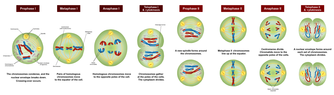
The stages of meiosis I and II. Image: Ali Zifan, CC BY-SA 4.0, via Wikimedia Commons.
🚀 NEET Advanced Edge
Distinguishing meiosis I vs meiosis II by cell appearance: In meiosis I, HOMOLOGOUS chromosomes (each still as a pair of sister chromatids, looking like an X) separate - anaphase I shows whole chromosomes moving to each pole. In meiosis II, SISTER chromatids separate - anaphase II looks just like mitotic anaphase but starting from a haploid cell. Exam questions often test this by showing a diagram and asking which division/stage it represents.
Why meiosis II resembles mitosis: Both separate sister chromatids; the key difference is meiosis II starts with a haploid (n) cell from meiosis I, while mitosis starts and ends diploid (2n) - same mechanical process, different starting ploidy.
Cancer connection: A mutated p53 ("guardian of the genome") fails to arrest the cell cycle or trigger apoptosis in cells with DNA damage, allowing damaged cells to keep dividing - this is why TP53 is the most commonly mutated gene across human cancers, a frequently tested NEET fact.
Regulation of the Cell Cycle
- Progression through the cell cycle is controlled at checkpoints by proteins called cyclins and enzymes called cyclin-dependent kinases (CDKs).
- A CDK becomes active only when it binds its specific cyclin; the resulting cyclin-CDK complex drives the cell from one phase to the next.
- Cyclin levels rise and fall at particular stages, so different cyclin-CDK complexes trigger the G1 to S and G2 to M transitions.
- The Restriction (R) point in G1 is the key decision point beyond which the cell is committed to divide.
- Faulty regulation and uncontrolled division of cells can lead to a mass of cells called a tumour, the basis of cancer.
Mitosis versus Meiosis: Key Differences
- Mitosis occurs in somatic (body) cells, whereas meiosis occurs in reproductive (germ) cells to form gametes/spores.
- Mitosis involves a single division producing two daughter cells; meiosis involves two successive divisions producing four daughter cells.
- Daughter cells of mitosis are diploid (2n) and genetically identical to the parent; those of meiosis are haploid (n) and genetically variable.
- Crossing over and pairing of homologous chromosomes (synapsis) occur only in meiosis (prophase I), not in mitosis.
- Mitosis maintains the chromosome number and helps growth and repair; meiosis halves the chromosome number, maintaining constancy across generations.
Karyokinesis and Cytokinesis
- Cell division (M phase) has two overlapping events: karyokinesis (division of the nucleus) followed by cytokinesis (division of the cytoplasm).
- Karyokinesis is divided into four stages: prophase, metaphase, anaphase and telophase.
- In animal cells, cytokinesis occurs by a cleavage furrow that constricts the cell from the outside inward.
- In plant cells, a rigid cell wall prevents furrowing, so a cell plate forms from the centre outward, giving rise to the new wall.
- Sometimes karyokinesis is not followed by cytokinesis, producing a multinucleate condition called a syncytium (e.g. liquid endosperm in coconut).
Significance and Applications of the Cell Cycle
- Growth of a multicellular organism from a single-celled zygote occurs through repeated cell division.
- Cell division continuously replaces old, dead and worn-out cells, for example cells of the skin and gut lining.
- The average duration of the human cell cycle is about 24 hours, with interphase occupying roughly 23 hours and M phase only about 1 hour.
- Some highly differentiated cells (e.g. mature neurons) exit the cycle into the inactive, non-dividing quiescent (G0) stage.